Right heart failure can be caused by persistent right ventricular overload which is a consequence of pulmonary hypertension. Many patients with primary pulmonary hypertension or chronic obstructive pulmonary disease (COPD) die of right heart failure. Pulmonary Artery Banding (PAB) models are very useful in developing effective therapy.
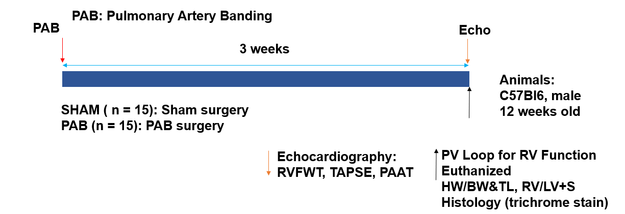
Experimental Protocol
The PAB model is induced surgically, and cardiac remodeling is monitored at specific timepoints during the study. A non-invasive echocardiogram is performed at 24 hours and 3 weeks post-PAB. A live pressure-volume loop is obtained at 3 weeks followed by final measurements, sample collection, and histology.
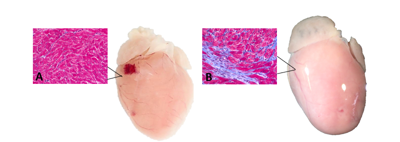
Histology
Figure 1. Cardiac remodeling and right ventricle fibrosis via Masson’s trichrome staining in PAB mice. (A) SHAM heart and histology. (B) PAB heart and histology.
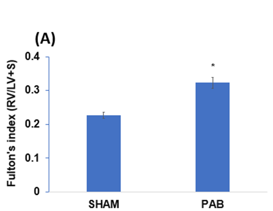
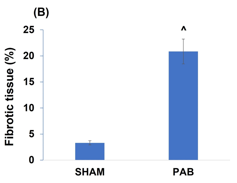
Right Ventricular Remodeling
Figure 2. (A) Cardiac remodeling in PAB mice, *: p <0.01, PAB vs. SHAM. (B) Cardiac fibrosis in PAB mice, ^: p<0.01, PAB vs. SHAM.
Mouse Echocardiography
Table 1. Echocardiogram at 24 hours and 3 weeks after
surgery in the SHAM and PAB groups.
Abbreviations: IVSd, Interventricular Septum Thickness at end Diastole; LVEDd, Left-Ventricular End-Diastolic Dimension; LVPTd, Left-Ventricle Posterior Wall Thickness Diastole; IVSs, Interventricular Septum Thickness at end Systole; LVESd, Left-Ventricular End-Systolic Dimension; LVPTs, Left-Ventricle Posterior Wall Thickness Systole; HR, Heart Rate; FS, Fractional Shortening; EF, Ejection Fraction; LVM, Left Ventricular Mass; RVFWT, Right Ventricle Free Wall Thickness; TAPSE, Tricuspid Annular Plane Systolic Excursion; PAAT, Pulmonary Artery Acceleration Time. All parameters are expressed as means ± the SD. The P value was calculated by comparing the final echo of the SHAM and PAB mice.
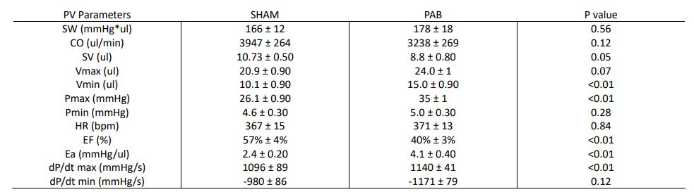
Right Ventricular Pressure-Volume Catheterization
Table 2. Right ventricular pressure-volume parameters (open-chest) in the SHAM and PAB groups.
Abbreviations: SW, Stroke Work; CO, Cardiac Output; SV, Stroke Volume; Vmax, Maximum Volume; Vmin, Minimum Volume; Pmax, Maximum Pressure; Pmin, Minimum Pressure; HR, Heart Rate; EF, Ejection Fraction; Ea, Arterial Elastance; dP/dt max, maximum derivative of change in systolic pressure over time; dP/dt min, minimum derivative of change in diastolic pressure over time.