The typical MI mouse study spans a duration of 4 weeks (28 days) as illustrated in the left panel. After 24 h post-surgery, mice were treated with vehicle (VE) or test article (TA).
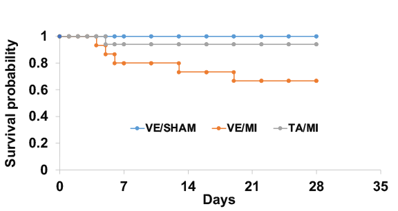
Survival Rate
Mice survival was monitored daily after MI. Survival rate was calculated during 30 days post-MI.
Heart Images
MI was induced in mice by permanently occluding the left coronary artery, performed under a stereo microscope. Each heart was photographed at the end of the experiment under the same multiplication.
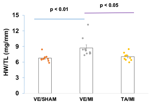
Cardiac Remodeling
The ratio of heart weight (HW) to tibia length (TL) was calculated to assess post-MI remodeling.
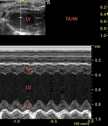
Mouse Echocardiography
Control Heart
Mouse heart B mode (upper panel) and M mode (lower panel).
Infarcted Heart
Left ventricle (LV) was dilated and inter ventricular septum (IVS) was decreased.
Treated Heart
The treatment attenuated LV dilation and IVS thinning.
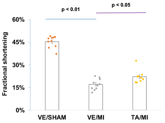
FS
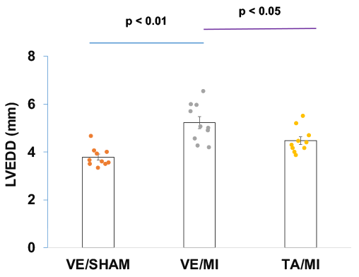
LVEDD
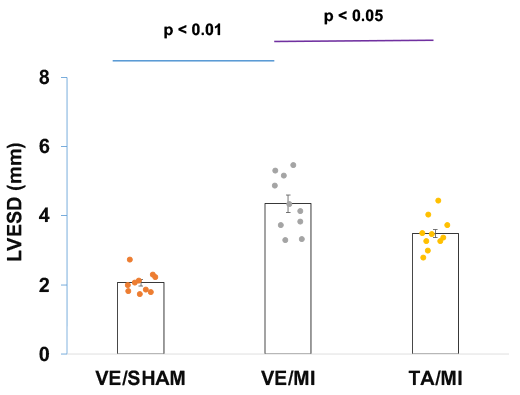
LVESD
In M mode, left ventricular end-diastole diameter (LVEDD) and end-systolic diameter (ESD) was measured and fractional shortening (FS) was calculated.
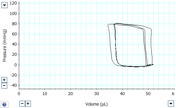
Mouse Pressure-Volume Catheterization
Normal Loop
Left ventricular pressure-volume (PV) was recorded simultaneously
Shrunken MI Loop
MI decreased LV pressure develop and chamber dilation
Improved MI Loop
LV contraction and dilation was improved after the treatment
CO
EF
+dp/dtmax
Cardiac output (left), ejection fraction (middle), and +dp/dtmax (right) were calculated from the recorded pressure and volume measurements.
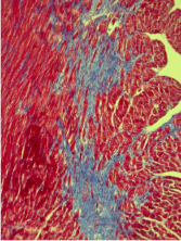
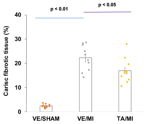
Masson's Trichrome Staining
VE/SHAM
Masson’s trichrome staining of cardiac section
VE/MI
Fibrotic tissue stained blue in the non-ischemic area
TA/MI
The treatment decreased cardiac fibrosis
Analysis
TA significantly reduced fibrotic tissue in the non-ischemic myocardium
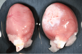
Rat MI Model
Rat MI model was generated by similar method. In the SHAM group (left), the left ventricular myocardium exhibited a consistent red appearance. In contrast, in the MI group (right), the infarcted myocardium transformed into white fibrotic tissue, indicating the presence of cardiac remodeling.