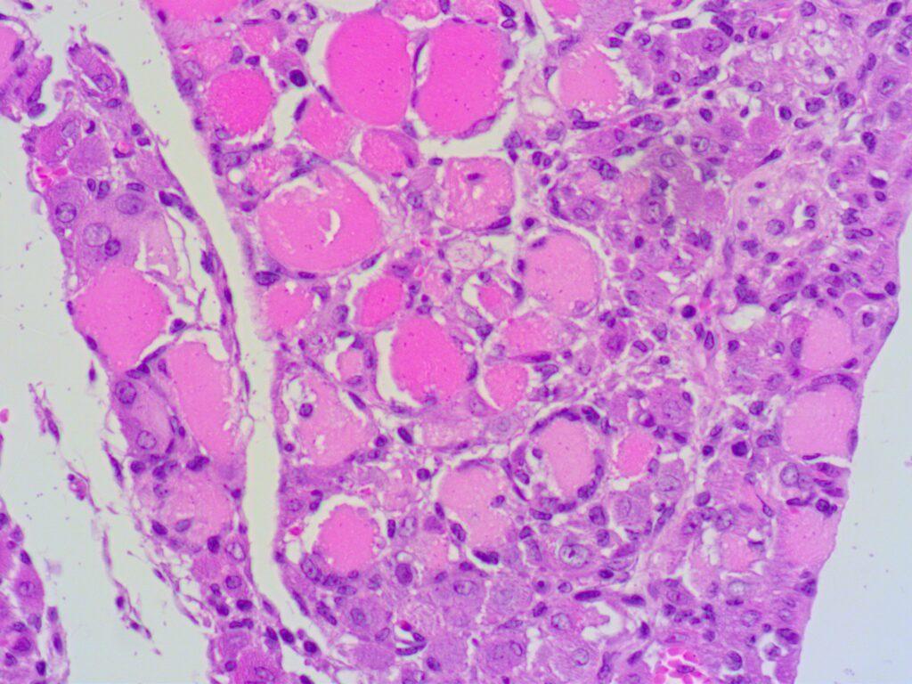
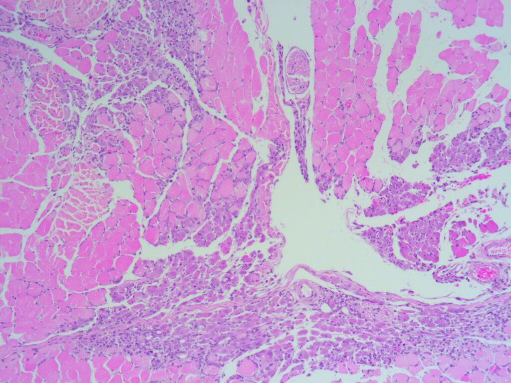
To surgically induce hindlimb ischemia, one-side common femoral artery and popliteal artery will be ligated and cut. The segment between the two arteries will be isolated and removed resulting in hindlimb ischemia. The blood flow (indicated by the color red in the figure below) is scanned using a Moor LDI laser image. There is no blood flow in the right hindlimb after the hindleg surgery (Left). One week later, H&E staining showed that loss of muscle mass and inflammatory cell infiltration (right, 400x)
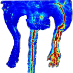
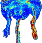
Blood Flow Recovery
After hindlimb ischemia, mice were treated with saline (left) and test article (right) for one week.
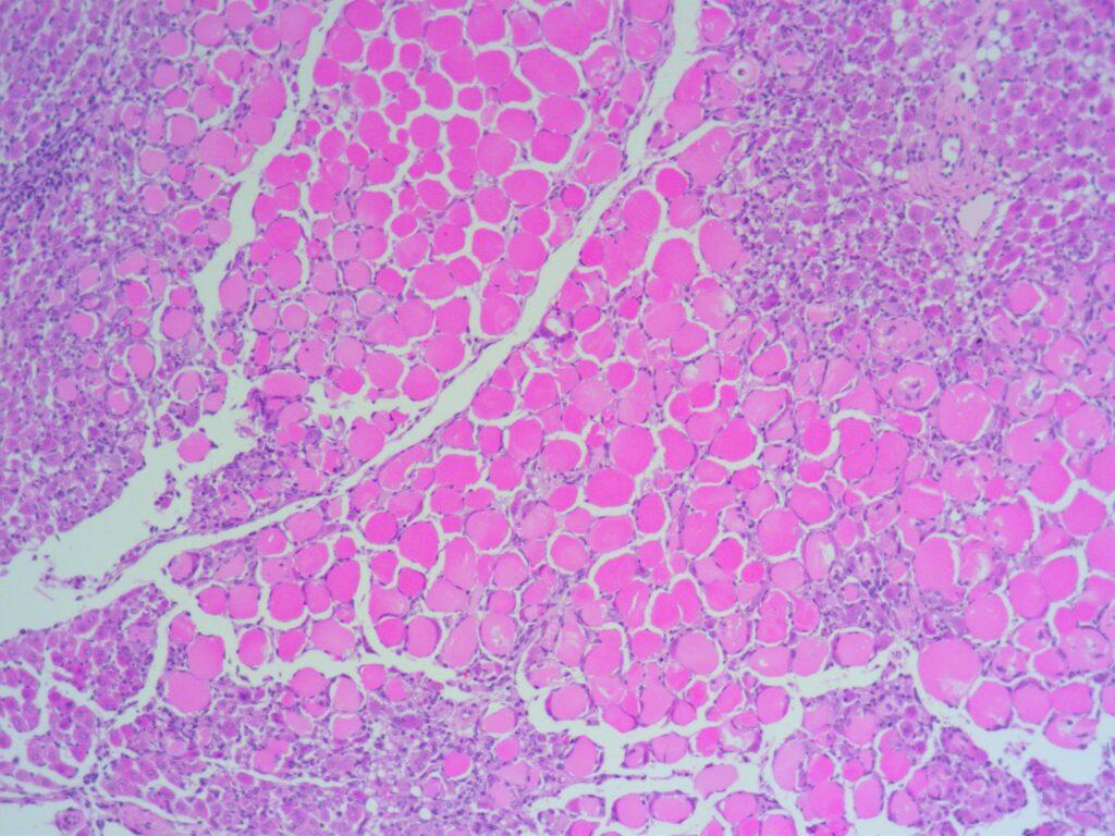
Tissue Injury
After hindlimb ischemia, mice were treated for one week. Tissue injury in control (left) and test article (right) mice was examined by H&E staining