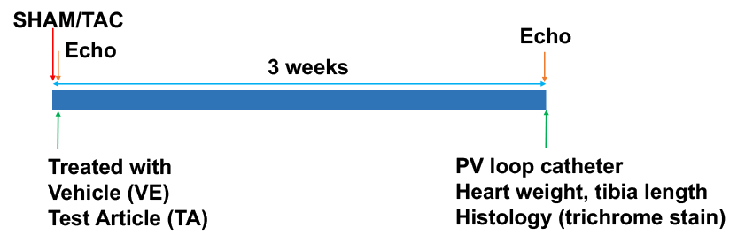
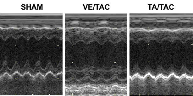
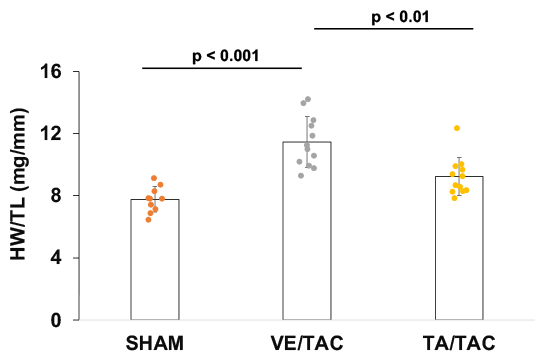
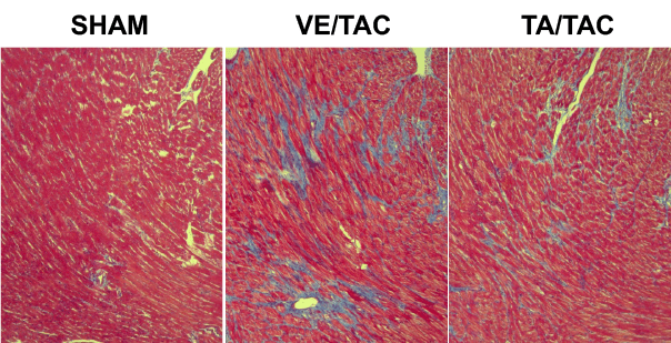
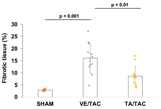
Mice received TAC or SHAM surgery. Echocardiography was performed on all mice at days 1 and 21 post-surgery. At the end of the experiments, left ventricular hemodynamics was assessed by a Millar pressure catheter. The ratio of heart weight (HW) to tibia length (TL) was measured in all mice. Cardiac sections were stained by Masson’s trichrome.
TA Improved Diastolic Function
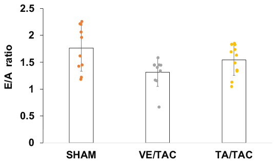
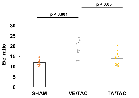
Echocardiography was performed on mice at the end of the experiments. TA inhibited left ventricular septal and posterior wall thickening (upper in parasternal short axis view). TA attenuated left ventricular E/A ratio reduction (lower left) and E/e’ ratio elevation (lower right)
TA Reduced Left Ventricular Contractility
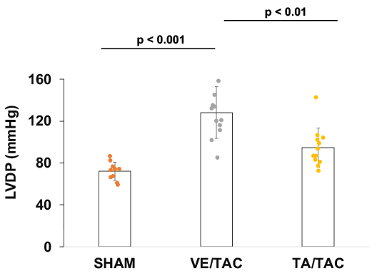
A Millar pressure catheter was inserted into the left ventricle at the end of the experiments. TA decreased left ventricular developed pressure (LVDP) (left) and +dp/dtmax (right) in TAC mice.
TA Decreased Cardiac Remodeling
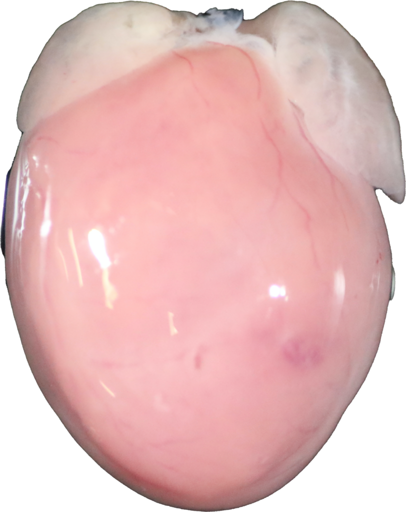
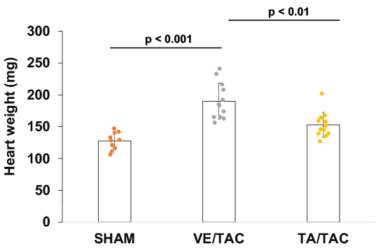
Hearts were photographed at the same amplification (upper). TA decreased heart weight (HW) (lower left) and HW to tibia length (TL) ratio (lower right) in TAC mice.
TA Inhibited Cardiac Fibrosis
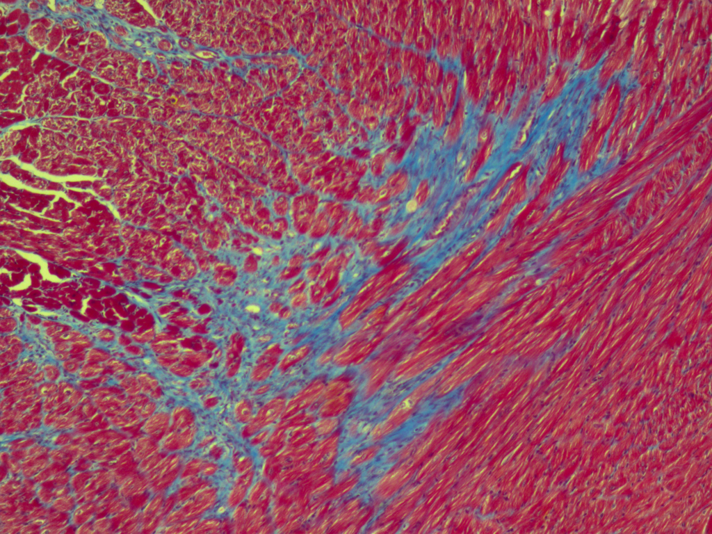
Cardiac sections were stained by Masson’s trichrome (upper). TA decreased cardiac fibrotic tissues in TAC mice (lower).