High fat diet induces obesity, type II diabetes, and cardiac hypertrophy. Administration of angiotensin II results in hypertension and worsens left ventricular remodeling in animals fed with high fat diet, leading to diastolic dysfunction.
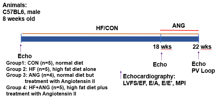
Experimental Protocol
C57BL6 mice started on a high fat diet, and they were were monitored for cardiac remodeling at specific timepoints during the study. After fed for 18 weeks, an Angiotensin II pump was surgically administered. A non-invasive echocardiogram was performed at the beginning, 18, and 22 weeks on the high fat diet. A live pressure-volume loop was obtained at 22 weeks followed by final measurements and sample collection.
Echocardiography
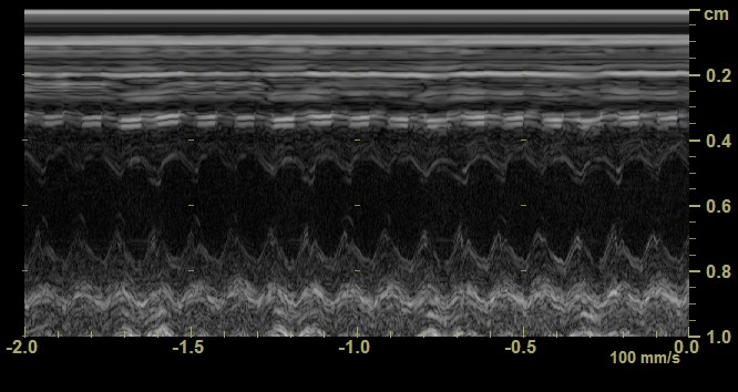
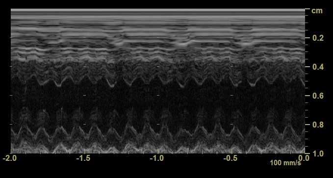
Figure 1. B-Mode echocardiography in the parasternal short-axis (PSAX) view of the left ventricle. The left panel depicts a C57BL6 mouse. The right panel depicts a high fat diet + angiotensin II mouse with 32% increase in the interventricular septum.
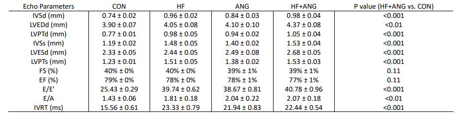
Table 1. Final echocardiogram parameters in CON, HF, ANG, and HF+ANG mice.
Abbreviations: CON, Control; HF, High Fat Diet; ANG, Angiotensin II; IVSd, Interventricular Septum Thickness at end Diastole; LVEDd, Left-Ventricular End-Diastolic Dimension; LVPTd, Left-Ventricle Posterior Wall Thickness Diastole; IVSs, Interventricular Septum Thickness at end Systole; LVESd, Left-Ventricular End-Systolic Dimension; LVPTs, Left-Ventricle Posterior Wall Thickness Systole; FS, Fractional Shortening; EF, Ejection Fraction; E/E’, Left Ventricular Diastolic Function; E/A, Left Ventricular Diastolic Function; IVRT, Isovolumetric Relaxation Time. All parameters are expressed as means ± the SEM. The P value was calculated by comparing the final echo between the HF+ANG and CON mice.
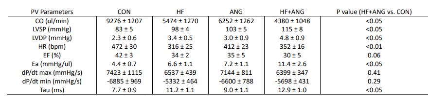
Table 2. Pressure-volume parameters of the left ventricle (open chest) in CON, HF, ANG, and HF+ANG mice.
Abbreviations: CON, Control; HF, High Fat Diet; ANG, Angiotensin II; CO, Cardiac Output; LVSP, Left Ventricle Systolic Pressure; LVDP, Left Ventricle Diastolic Pressure; HR, Heart Rate; EF, Ejection Fraction; Ea, Arterial Elastance; dP/dt max, maximum derivative of change in systolic pressure over time; dP/dt min, minimum derivative of change in diastolic pressure over time; Tau, Left Ventricle Relaxation Constant.
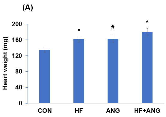
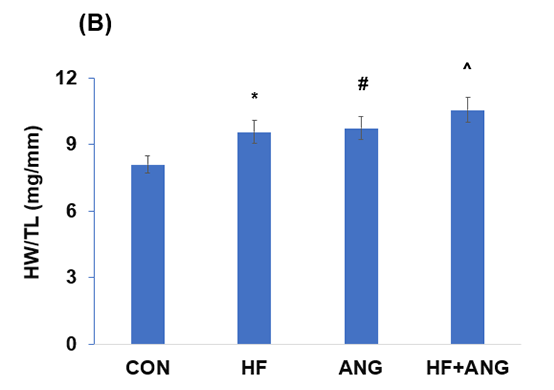
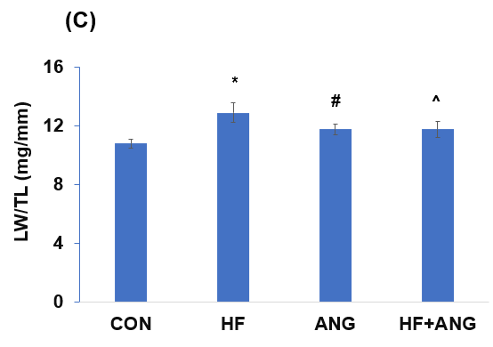
Cardiac and Pulmonary Remodeling
Figure 2. Cardiac and pulmonary remodeling. (A) High fat diet (HF) and angiotensin II (ANG) induced cardiac hypertrophy demonstrated by heart weight in control (CON), HF, ANG, and HF+ANG groups. *: p<0.05, HF vs. CON. #: p<0.05, ANG vs. CON. ^: p<0.01, HF+ANG vs. CON. (B) HF and ANG induced cardiac hypertrophy demonstrated by heart weight/tibia length in CON, HF, ANG, and HF+ANG groups. *: p<0.05, HF vs. CON. #: p<0.05, ANG vs. CON. ^: p<0.01, HF+ANG vs. CON. (C) HF induced pulmonary remodeling demonstrated by lung weight/tibia length in CON, HF, ANG, and HF+ANG groups. *: p<0.05, HF vs. CON. #: p=0.11, ANG vs. CON. ^: p=0.15, HF+ANG vs. CON.