Pressure-Volume Loop Catheterization
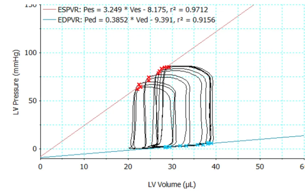
Figure 1. Left-ventricular end-systolic and end-diastolic pressure-volume relationship of a SHAM mouse after performing an occlusion of the inferior vena cava.
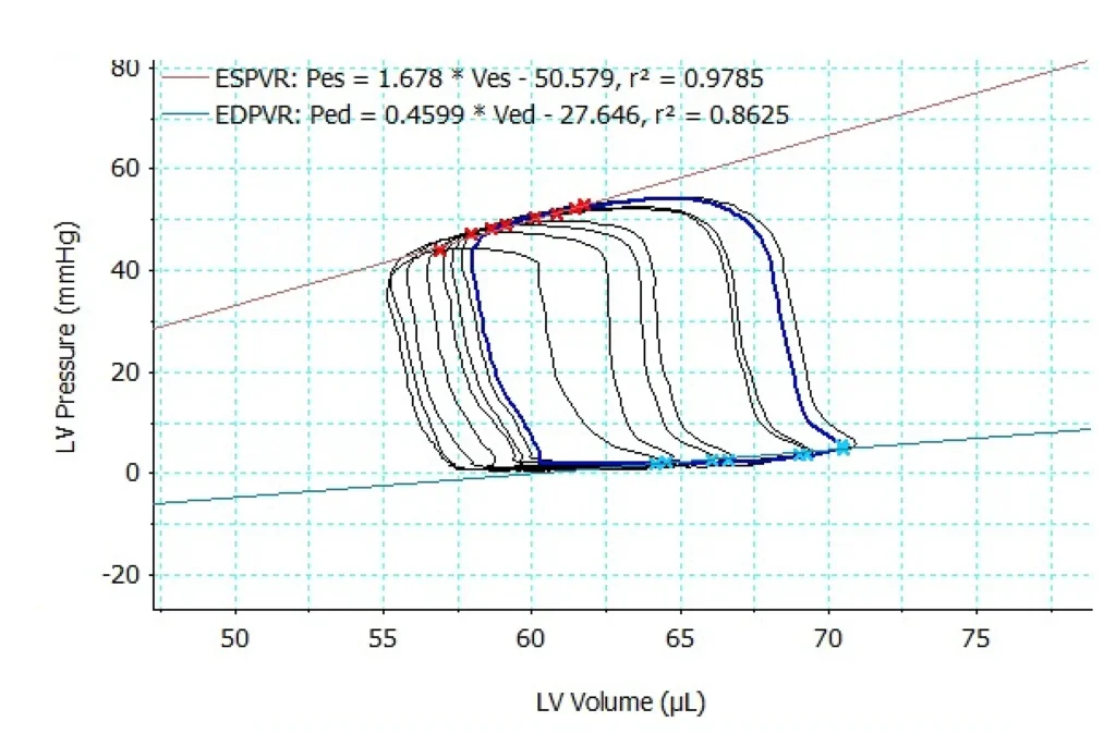
Figure 2. Left-ventricular end-systolic and end-diastolic pressure-volume relationship of a MI animal model.
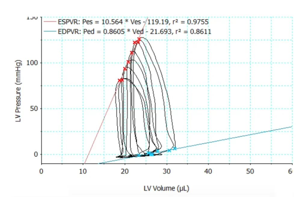
Figure 3. Left-ventricular end-systolic and end-diastolic pressure-volume relationship of a TAC animal model.
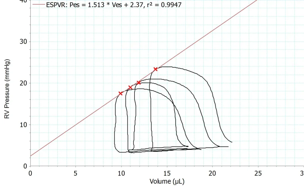
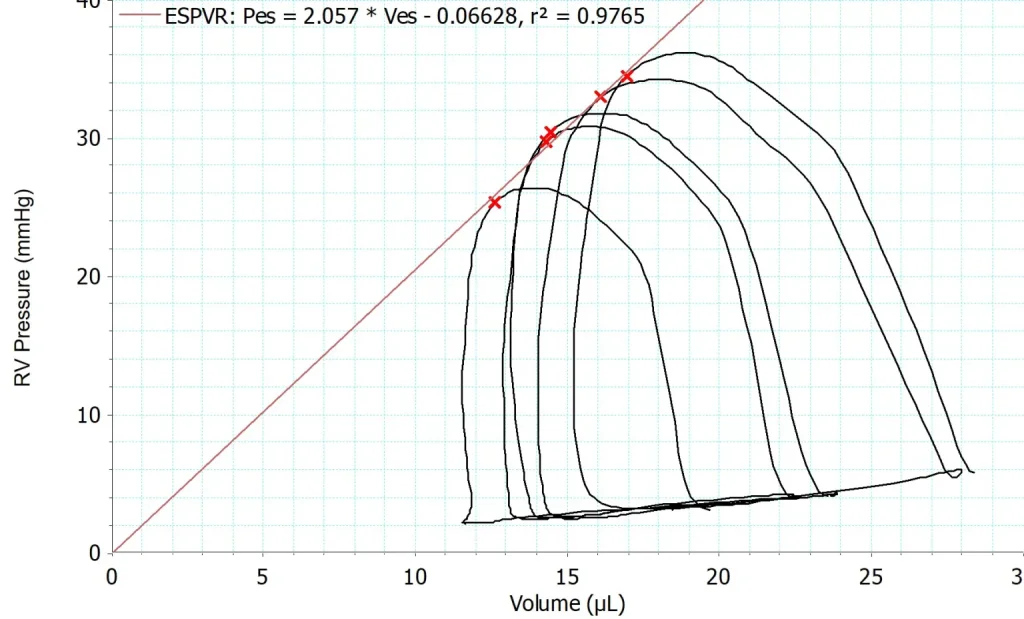
Figure 4. The left pressure-volume loop shows the end-systolic pressure-volume relationship of the right ventricle of a SHAM mouse. The right pressure-volume loop shows the end-systolic pressure-volume relationship of the right ventricle of a mouse that underwent pulmonary artery banding (PAB) surgery.
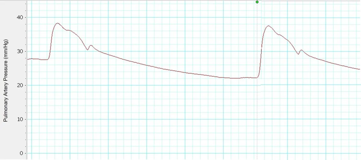
Figure 5. The pulmonary artery pressure of a PAB animal model.
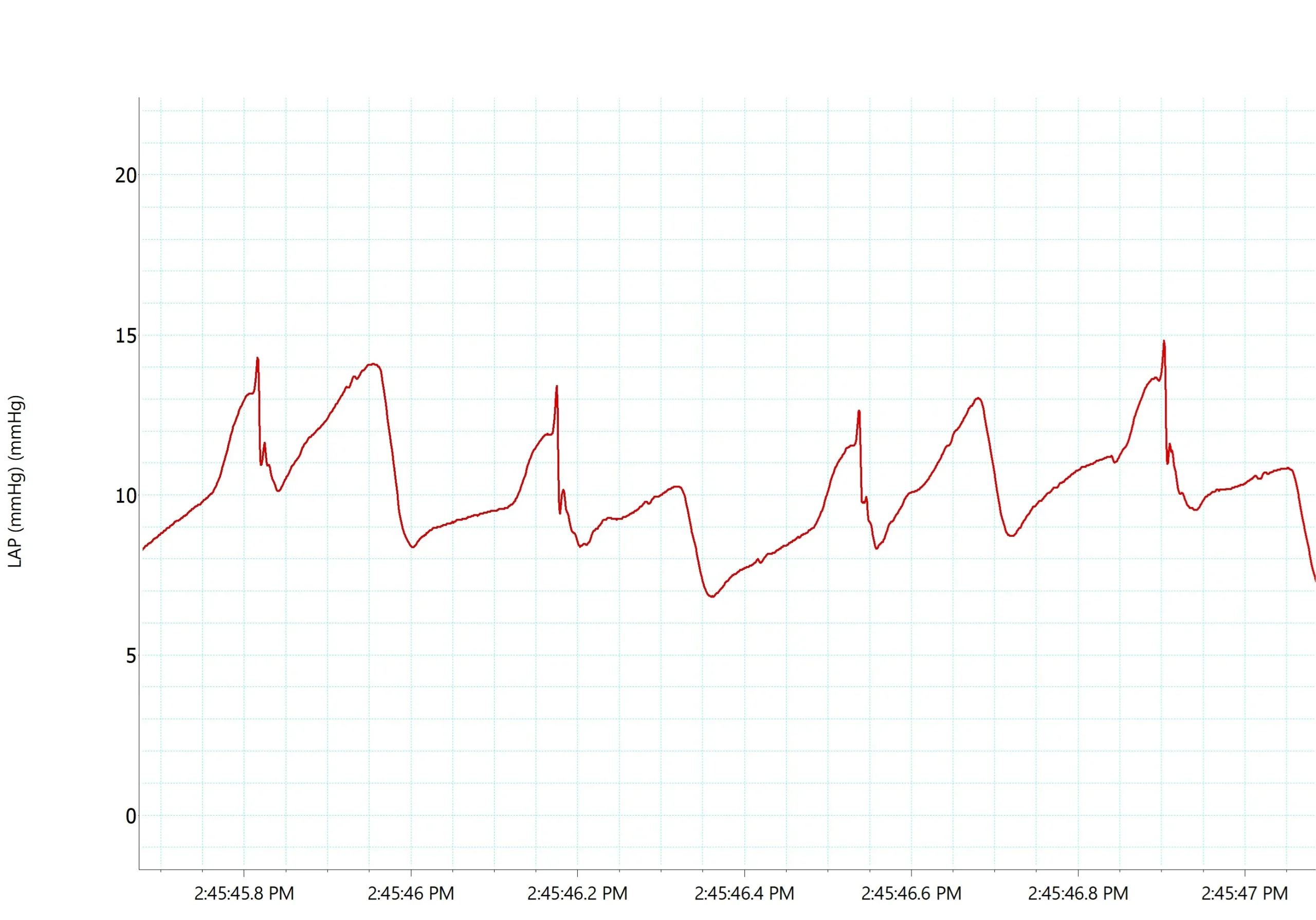
Figure 6. The left atrial pressure of a ZSF1 diabetic rat.


